When you elevate your heel, your calf muscles enable you to move forward. If you didn’t have those muscles, you wouldn’t be able to move at all. The cadence and explosiveness of your stride are enhanced by strong calves.
by Dr. Neeraj Mehta (Professor at American Sports Fitness University & COO of GFFI Fitness Academy)
The function of calf muscles:
When the foot comes into contact with the ground, the calf muscles slow down the bending and straightening forces applied to the knee and ankle joints.
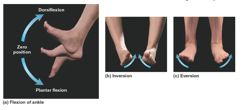
Gluteal muscle (soleus) is stretched when the heel touches the ground during a stride or lunge, or when the knee bends when lowering oneself to sit down (dorsiflexion).
The gastrocnemius extends when the knee straightens during flexion of the leg behind the hip. Instead of loosening up as they stretch, these muscles extend under stress as they work to slow down the forces acting on the knee and ankle joints during flexion and extension.
Here is a look into Calf’s structure.
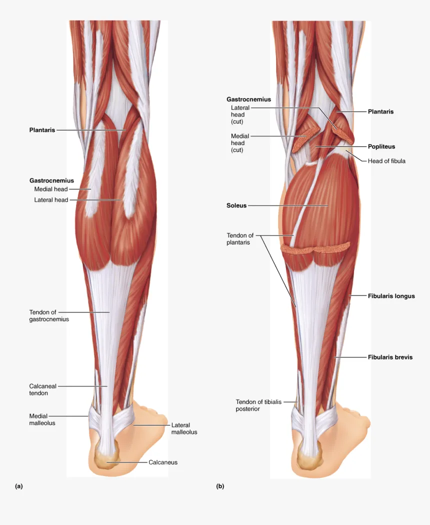
Gastrocnemius
In order to walk and maintain an upright posture, the gastrocnemius muscle is a vital part of the body. Gastrocnemius is a robust muscle that constitutes the majority of the rear of the lower thigh. From the back of the knee to the heel, it is a two-joint or biarticular muscle with two heads. The medial and lateral heads of the gastrocnemius, located near the back of the lower leg’s upper half, give the calf its distinctive form.
Soleus
Found in the leg’s superficial posterior compartment You may find the Soleus muscle deep within of the gastronemius muscle in your lower leg.
Plantaris
The Triceps Surae is made up of the Plantaris, Gastrocnemius, and Soleus muscles, all of which have short bellies and long, thin tendons. They are all positioned in the leg’s posterior compartment.
Lever consideration of Calf Muscles
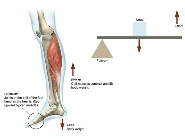
However, the gastrocnemius in your calf has a mechanical advantage. The lower leg operates as a second class lever during plantar flexion. The only lever of second class that guarantees that the effort arm will always be larger than the load arm is a second class lever. As a consequence of this configuration, the effort arm to load arm ratio is increased, making the second class lever the most mechanically beneficial.
The gastrocnemius muscle, which is linked to the calcaneus bone, provides the effort for calf raises. The load is generated by your body weight and the additional weight you are carrying; this force is transmitted to the lever system through the tibia. The metacarpophalengeal joint serves as the fulcrum.
The load is centered in this design, while the effort is positioned furthest from the fulcrum. Thus, even if your bicep is as powerful as your calf, the act of plantarflexion may transfer far more weight than elbow flexion.
When your calves are weak or tight, you are more prone to a variety of problems, including shin splints. Keeping your calf muscles in excellent shape by strengthening them on a regular basis, as well as stretching and foam rolling, can help you avoid injury.
Biomechanics of the Calf Movement / Exercise
Performing calf raises engages the gastrocnemius, back of knee, and soleus muscles at the bottom of the foot. The movement is known as plantar flexion, or ankle extension.
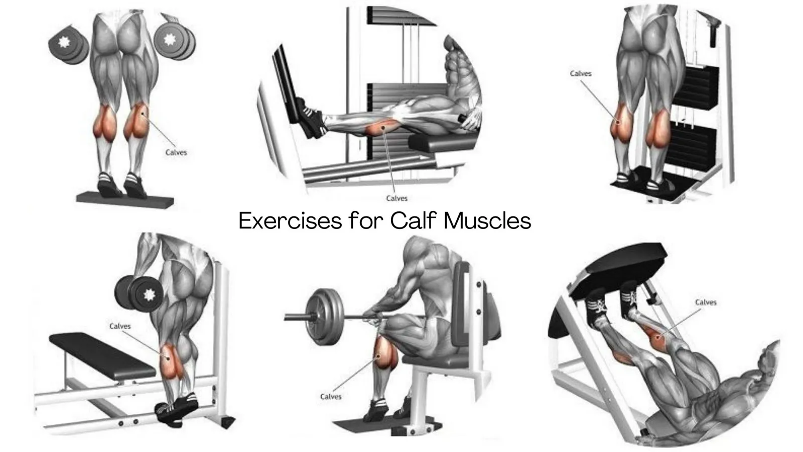
We can better understand why the calf raise isn’t working by studying biomechanics and physics. The calf muscle acts as a spring tensioner for the Achilles tendon. When the calf contracts, it braces against the back of the shin bone and the back of the knee. Because each muscle fiber lines up at an angle and does not line up vertically, the surrounding tissue is yanked obliquely rather than longitudinally, making this calf’s structure ideal for this purpose. The surrounding tissue’s stress creates a spring in the Achilles tendon, allowing it to propel you forward or upward.
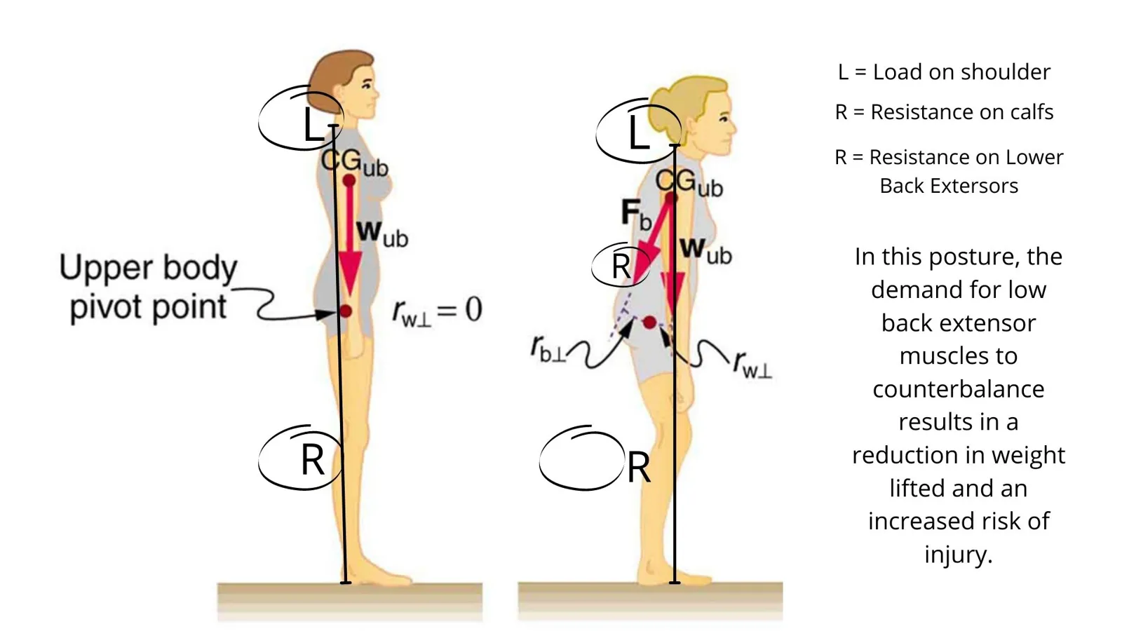
To put it simply, the two fundamental biomechanical issues that arise from incorrectly completing this exercise are caused by leaning forward and extending the knees.
First, a moment arm is formed in the low back by the vertical force line extending from the point of contact between the shoulders and the weight. The low back extensor muscles will be required to counterbalance this flexion moment.
The knee hyperextension moment is the second biomechanical consequence of this bad workout method. Because the force line passes anterior to the knees, hyperextension ensues.
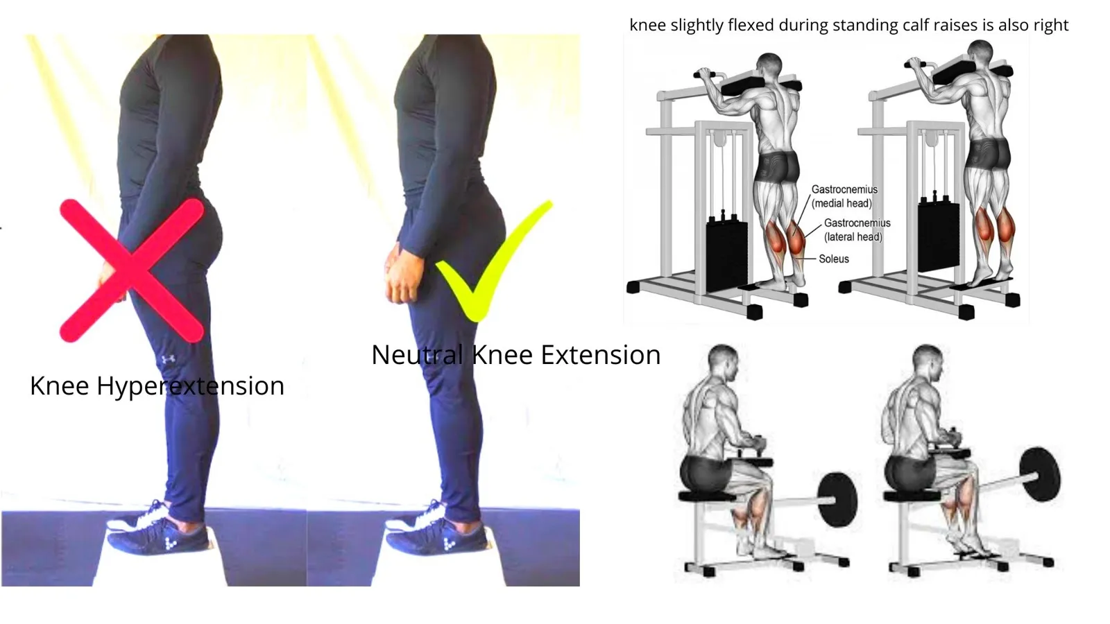
Maximum hyperextension of the knees in men is 1.0°, whereas that of the knees in females is 1.6°.
Soft tissues like ligaments serve as the body’s first line of defense against damage because of the significant hyperextension moment experienced by the “locked” knee in this posture.
Maintaining a neutral spine and slightly flexed knees when completing a standing calf raise is essential for appropriate biomechanics.
Seated Calf Raises Requirement
The soleus muscle is the only one that seated calf raises can effectively target, hence they should be included in every strengthening routine. When an athlete moves from full stretch to full contraction with a bent knee, they enhance their dorsiflexion ability (in terms of range of motion) as well as their ground response force during sprints and leaps (in terms of vertical jump). To assist avoid injuries, it will also strengthen the tibia’s capacity for posterior translation. An essential consideration is whether or if an athlete has previously been doing standing calf raises, which target the gastrocnemius, an antagonist of the anterior knee ligament (ACL). The chance of problems rises if you do not train your muscles in a balanced manner.
Participation in Other Activities
When the ankles are dorsally flexed, the Gastrocnemius may aid in knee flexion (ie: Leg Curl). Squats and other complex Quadriceps movements, in which the knee is moved forward from the ankle, also use the plantar flexors. Moving over both ankle and knee joints at the same time, the Gastrocnemius functions as a Dynamic Stabilizer, while the Soleus acts as a Synergist by dorsal flexing the ankle. The plantar flexors also play a role in lower body plyometrics and Olympic weightlifting, which require hopping.
A runner and a calf Muscle
Running relies heavily on the calf muscles, which provide a wide range of functions: During the swing phase, the tibialis anterior, a muscle running along the front of the shin, acts as a rocker to raise the heels off the ground. To urge you forward, it places your weight on your toes.
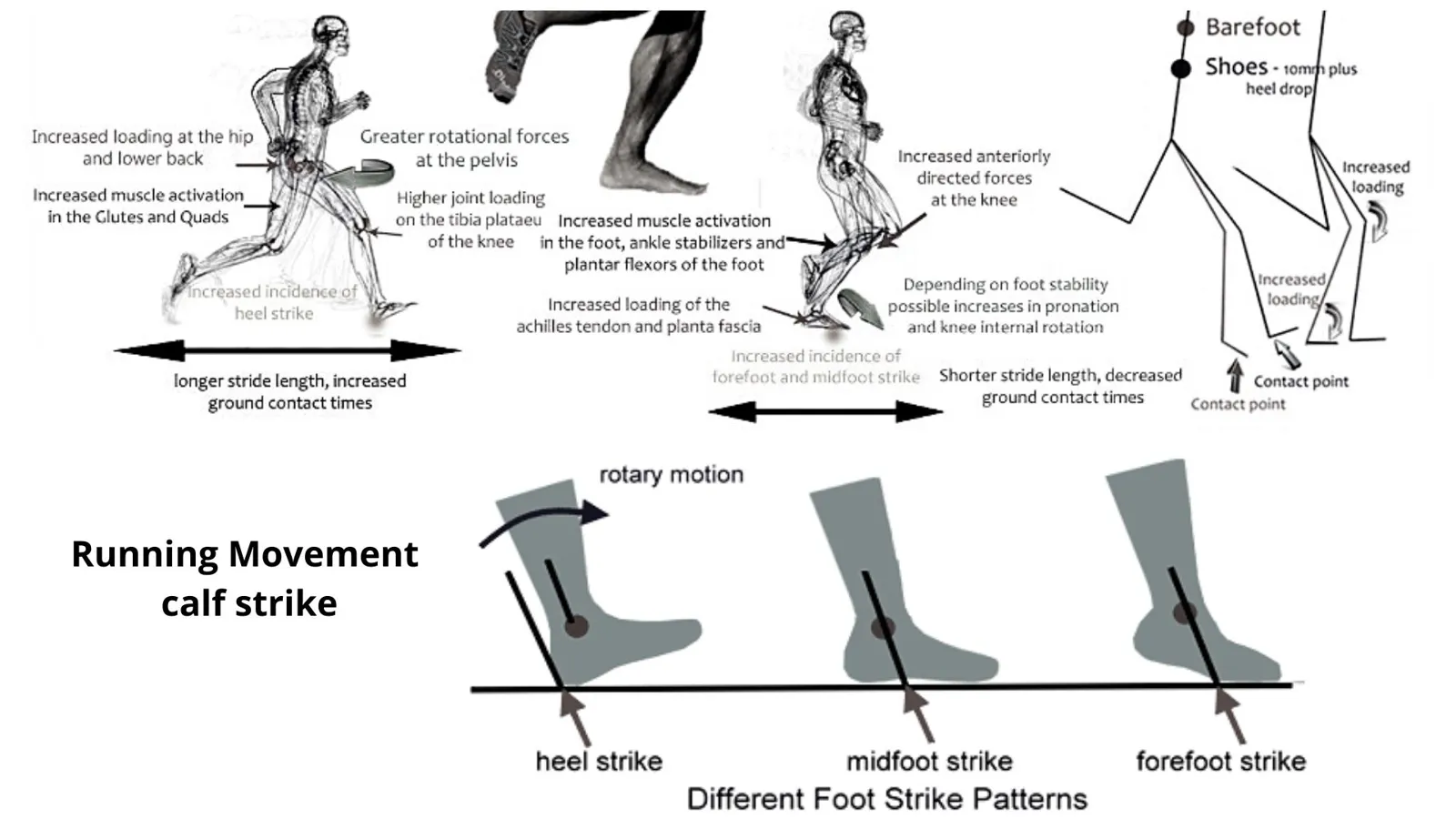
In the second part of the running stride, the calf muscles are most engaged (foot on the ground). They exert eccentric (controlled lengthening) control over the tibia’s forward translation (to prevent you from falling on your face). In late midstance to terminal stance, the calf muscles swiftly stretch (plantarflex) the ankle, pushing the body forward concentrically. Forefoot and midfoot landing patterns may be prepared for toward the conclusion of the swing phase (airborne) of the running stride, which is rather silent until then.
A recent Finnish research discovered that when runners were connected to 3-D motion capture devices, the total effort needed of the calf muscles was actually 25% larger than that required of the quadriceps. This makes them more susceptible to exhaustion during a lengthy run or race, thereby slowing your speed.
Strengthening the calf muscles of runners is a need.
Strengthening the Calf Muscle is essential for runners. Calf muscles are responsible for a substantial portion of the body’s propulsion when running, thus it seems logical that improved strength and power production might also lead to quicker running! As a result, maintaining good condition of these muscles is critical from both an injury prevention and performance perspective.
It’s one of the best workouts you can perform to improve your running. Training the calf, quad and hip as well as working on pelvic and trunk stability was accomplished through fast repeated single leg hops.
Calf muscle-related injuries
A frequent running injury is a strained calf muscle, which may be painful and debilitating because of the time it takes for it to recover and the likelihood that it will happen again.
Extrinsic and intrinsic elements have a role. In most cases, the calf will be exposed to external variables such as an unexpected load or a load that it is not trained to bear.
If your calf isn’t properly prepared, even practicing plyometrics, hill running, speed training, or running greater distances might cause harm.
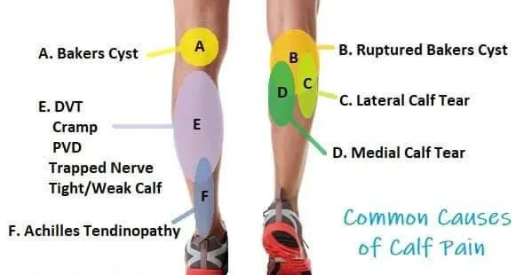
An injury is more probable when the calf is stretched while also being forced to contract hard and fast. This results in trauma. Both the gastrocnemius and soleus muscles in the calves are inflexible, and overpronating or oversupinating feet are also typical reasons because they disrupt the effective mechanical function of the foot and therefore increase the pressure on the calves, which may lead to pain.
A sciatic nerve impingement is another fairly prevalent reason. The sciatic nerve travels from the back of the leg to the foot, passing via the hamstring and calf muscles. Your back, pelvis, or even your hamstrings might cause it to become inflamed.
Protective spasms of the hamstring and calf muscles may occur in certain situations, which can make these muscles more vulnerable to injury.
When the muscle is loaded excessively strongly, it might induce an acute spasm that causes discomfort in the muscle or it can impair the muscle’s capacity to contract and relax, resulting in tearing.
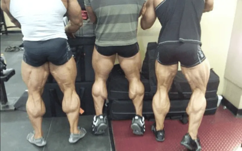
So what are you waiting for!!! work for your calfs to built overall strength
Important Notes:
- It’s important to keep your ankles and feet in good shape by working out your Soleus and Gastrocnemius (calf muscles).
- Must include Seated calf raises to strength calfs muscles.
- Weak or tight calves are more prone to a variety of problems, including shin splints
Reference:
https://schoolofgainz.com/calf-training/
https://www.physio-pedia.com/Gastrocnemius
https://www.medicalnewstoday.com/articles/lower-leg-anatomy
https://pilotfeasibilitystudies.biomedcentral.com/articles/10.1186/s40814-020-00639-5
https://exrx.net/Kinesiology/CalfExercises
https://www.openfit.com/calves-workout-exercises
https://greatist.com/fitness/calf-exercises
https://www.ncbi.nlm.nih.gov/pmc/articles/PMC5583613/
https://www.sciencedirect.com/science/article/pii/S0741521417300812
https://sportsmedicine-open.springeropen.com/articles/10.1186/s40798-021-00364-0
https://www.sspc.com.au/wp-content/uploads/2020/07/Green__Pizzari_2017_-Calf_Muscle_Strain_Injuries_in_Sport.pdf https://journals.sagepub.com/doi/abs/10.1177/0363546520959327 https://sma.org.au/resources-advice/injury-fact-sheets/gastrocnemius-calf-strain/
जब आप अपनी एड़ी ऊपर उठाते हैं, तो पिंडली की मांसपेशियाँ (calf muscles) आपको आगे बढ़ने में सक्षम बनाती हैं। यदि ये मांसपेशियाँ न होतीं, तो आप बिल्कुल भी गति नहीं कर पाते। सुदृढ़ पिंडलियाँ आपके कदमों की लय (cadence) और विस्फोटकता को बढ़ाती हैं।
डॉ. नीरज मेहता, PhD द्वारा (American Sports Fitness University में प्रोफेसर एवं GFFI Fitness Academy के COO)
पिंडली की मांसपेशियों का कार्य:
जब पैर भूमि के संपर्क में आता है, तब पिंडली की मांसपेशियाँ घुटने और टखने के जोड़ों पर लगने वाले मोड़ने और सीधा करने वाले बलों की गति को मंद करती हैं।
चलते समय कदम या लंज (lunge) के दौरान जब एड़ी भूमि को स्पर्श करती है, अथवा बैठने के लिए नीचे आते समय जब घुटना मुड़ता है (डॉर्सीफ्लेक्शन — dorsiflexion), तब ग्लूटियल मांसपेशी (सोलियस — soleus) खिंचती है।
कूल्हे के पीछे पैर के फ्लेक्शन के दौरान जब घुटना सीधा होता है, तब गैस्ट्रोक्नीमियस (gastrocnemius) लंबी होती है। खिंचाव के साथ ढीली पड़ने के स्थान पर, ये मांसपेशियाँ तनाव के अधीन लंबी होती हैं, क्योंकि वे फ्लेक्शन और एक्सटेंशन के दौरान घुटने और टखने के जोड़ों पर कार्य करने वाले बलों को मंद करने का कार्य करती हैं।
यहाँ पिंडली की संरचना पर एक दृष्टि है।
गैस्ट्रोक्नीमियस (Gastrocnemius)
चलने और सीधी मुद्रा बनाए रखने के लिए गैस्ट्रोक्नीमियस मांसपेशी शरीर का एक अत्यावश्यक अंग है। गैस्ट्रोक्नीमियस एक सशक्त मांसपेशी है जो निचली जाँघ के पिछले भाग का अधिकांश हिस्सा बनाती है। घुटने के पीछे से एड़ी तक फैली यह दो सिरों वाली, दो-संधि अथवा द्वि-संधिक (biarticular) मांसपेशी है। निचले पैर के ऊपरी आधे भाग के पीछे स्थित गैस्ट्रोक्नीमियस के मध्य (medial) और पार्श्व (lateral) सिर पिंडली को उसका विशिष्ट आकार प्रदान करते हैं।
सोलियस (Soleus)
यह पैर के सतही पश्च कक्ष (superficial posterior compartment) में पाई जाती है। सोलियस मांसपेशी आपके निचले पैर में गैस्ट्रोक्नीमियस मांसपेशी के भीतर गहराई में स्थित होती है।
प्लांटेरिस (Plantaris)
ट्राइसेप्स सुरी (Triceps Surae) प्लांटेरिस, गैस्ट्रोक्नीमियस और सोलियस मांसपेशियों से मिलकर बनती है, जिन सभी के पेट (bellies) छोटे और टेंडन लंबे तथा पतले होते हैं। ये सभी पैर के पश्च कक्ष (posterior compartment) में स्थित होती हैं।
पिंडली की मांसपेशियों का लीवर विश्लेषण
तथापि, आपकी पिंडली की गैस्ट्रोक्नीमियस को यांत्रिक लाभ (mechanical advantage) प्राप्त है। प्लांटर फ्लेक्शन (plantar flexion) के दौरान निचला पैर द्वितीय श्रेणी के लीवर (second class lever) के रूप में कार्य करता है। द्वितीय श्रेणी का लीवर ही एकमात्र ऐसा लीवर है जो यह सुनिश्चित करता है कि प्रयास भुजा (effort arm) सदैव भार भुजा (load arm) से बड़ी रहेगी। इस विन्यास के परिणामस्वरूप प्रयास भुजा और भार भुजा का अनुपात बढ़ जाता है, जिससे द्वितीय श्रेणी का लीवर यांत्रिक रूप से सर्वाधिक लाभकारी बनता है।
कैल्केनियस (calcaneus) अस्थि से जुड़ी गैस्ट्रोक्नीमियस मांसपेशी काफ़ रेज़ (calf raises) के लिए प्रयास प्रदान करती है। भार आपके शरीर के वज़न और आपके द्वारा उठाए गए अतिरिक्त वज़न से उत्पन्न होता है; यह बल टिबिया (tibia) के माध्यम से लीवर तंत्र तक संचरित होता है। मेटाकार्पोफैलेंजियल जोड़ (metacarpophalengeal joint) आलंब (fulcrum) का कार्य करता है।
इस संरचना में भार मध्य में स्थित होता है, जबकि प्रयास आलंब से सबसे दूर स्थित होता है। इस प्रकार, भले ही आपका बाइसेप उतना ही शक्तिशाली हो जितनी आपकी पिंडली, प्लांटरफ्लेक्शन की क्रिया एल्बो फ्लेक्शन की तुलना में कहीं अधिक भार स्थानांतरित कर सकती है।
जब आपकी पिंडलियाँ दुर्बल या जकड़ी हुई होती हैं, तो आप शिन स्प्लिंट्स (shin splints) सहित अनेक समस्याओं के प्रति अधिक प्रवण होते हैं। पिंडली की मांसपेशियों को नियमित रूप से सुदृढ़ करके, तथा स्ट्रेचिंग और फोम रोलिंग के माध्यम से उन्हें उत्कृष्ट स्थिति में रखने से चोट से बचाव में सहायता मिल सकती है।
पिंडली की गति / व्यायाम की बायोमैकेनिक्स
काफ़ रेज़ करने से गैस्ट्रोक्नीमियस, घुटने का पिछला भाग, और पैर के निचले भाग की सोलियस मांसपेशियाँ सक्रिय होती हैं। इस गति को प्लांटर फ्लेक्शन, अथवा टखने का एक्सटेंशन कहा जाता है।
बायोमैकेनिक्स और भौतिकी के अध्ययन द्वारा हम बेहतर समझ सकते हैं कि काफ़ रेज़ प्रभावी क्यों नहीं हो रहा है। पिंडली की मांसपेशी एकिलीज़ टेंडन (Achilles tendon) के लिए स्प्रिंग टेंशनर का कार्य करती है। जब पिंडली संकुचित होती है, तो वह पिंडली की अस्थि (shin bone) के पिछले भाग और घुटने के पिछले भाग के विरुद्ध टिकती है। चूँकि प्रत्येक मांसपेशी तंतु ऊर्ध्वाधर रूप से न होकर एक कोण पर संरेखित होता है, इसलिए आसपास का ऊतक अनुदैर्ध्य रूप से नहीं बल्कि तिरछे खिंचता है, जिससे पिंडली की यह संरचना इस प्रयोजन के लिए आदर्श बनती है। आसपास के ऊतक का तनाव एकिलीज़ टेंडन में एक स्प्रिंग उत्पन्न करता है, जिससे वह आपको आगे या ऊपर की ओर प्रक्षेपित कर पाता है।
सरल शब्दों में, इस व्यायाम को गलत ढंग से करने से उत्पन्न होने वाली दो मूलभूत बायोमैकेनिकल समस्याएँ आगे झुकने और घुटनों को पूरी तरह सीधा करने के कारण होती हैं।
पहली, कंधों और भार के संपर्क-बिंदु से निकलने वाली ऊर्ध्वाधर बल-रेखा द्वारा कमर के निचले भाग में एक मोमेंट आर्म (moment arm) बनता है। इस फ्लेक्शन मोमेंट को संतुलित करने के लिए कमर की एक्सटेंसर मांसपेशियों को कार्य करना पड़ेगा।
इस त्रुटिपूर्ण व्यायाम पद्धति का दूसरा बायोमैकेनिकल परिणाम घुटने का हाइपरएक्सटेंशन मोमेंट (hyperextension moment) है। चूँकि बल-रेखा घुटनों के आगे से गुज़रती है, इसलिए हाइपरएक्सटेंशन उत्पन्न होता है।
पुरुषों में घुटनों का अधिकतम हाइपरएक्सटेंशन 1.0° होता है, जबकि महिलाओं में यह 1.6° होता है।
इस मुद्रा में “लॉक” घुटने पर पड़ने वाले उल्लेखनीय हाइपरएक्सटेंशन मोमेंट के कारण, लिगामेंट जैसे कोमल ऊतक क्षति के विरुद्ध शरीर की रक्षा की पहली पंक्ति का कार्य करते हैं।
स्टैंडिंग काफ़ रेज़ करते समय उचित बायोमैकेनिक्स के लिए तटस्थ रीढ़ (neutral spine) और हल्के मुड़े हुए घुटने बनाए रखना अनिवार्य है।
सीटेड काफ़ रेज़ की आवश्यकता
सीटेड काफ़ रेज़ (seated calf raises) ही एकमात्र ऐसा व्यायाम है जो सोलियस मांसपेशी को प्रभावी रूप से लक्षित कर सकता है, इसलिए इसे प्रत्येक सुदृढ़ीकरण दिनचर्या में सम्मिलित किया जाना चाहिए। जब कोई एथलीट मुड़े हुए घुटने के साथ पूर्ण खिंचाव से पूर्ण संकुचन तक गति करता है, तो वह अपनी डॉर्सीफ्लेक्शन क्षमता (गति-सीमा के संदर्भ में) तथा स्प्रिंट और छलाँगों के दौरान अपने भू-प्रतिक्रिया बल (ground response force) (ऊर्ध्वाधर छलाँग के संदर्भ में) — दोनों को बढ़ाता है। चोटों से बचाव में सहायता के लिए यह टिबिया की पश्च स्थानांतरण (posterior translation) क्षमता को भी सुदृढ़ करेगा। एक महत्वपूर्ण विचारणीय बिंदु यह है कि क्या एथलीट पहले से स्टैंडिंग काफ़ रेज़ कर रहा है, जो अग्र घुटना लिगामेंट (ACL) की प्रतिपक्षी (antagonist) गैस्ट्रोक्नीमियस को लक्षित करता है। यदि आप अपनी मांसपेशियों को संतुलित ढंग से प्रशिक्षित नहीं करते, तो समस्याओं की संभावना बढ़ जाती है।
अन्य गतिविधियों में सहभागिता
जब टखने डॉर्सली फ्लेक्स्ड होते हैं, तब गैस्ट्रोक्नीमियस घुटने के फ्लेक्शन (अर्थात् लेग कर्ल — Leg Curl) में सहायता कर सकती है। स्क्वाट और क्वाड्रिसेप्स की अन्य जटिल गतियाँ, जिनमें घुटना टखने से आगे की ओर ले जाया जाता है, भी प्लांटर फ्लेक्सर्स का उपयोग करती हैं। टखने और घुटने — दोनों जोड़ों पर एक साथ गति करते हुए गैस्ट्रोक्नीमियस गतिशील स्थिरक (Dynamic Stabilizer) के रूप में कार्य करती है, जबकि सोलियस टखने को डॉर्सली फ्लेक्स करके सहक्रियक (Synergist) के रूप में कार्य करती है। प्लांटर फ्लेक्सर्स निचले शरीर के प्लायोमेट्रिक्स और ओलंपिक वेटलिफ्टिंग में भी भूमिका निभाते हैं, जिनमें उछलना आवश्यक होता है।
धावक और पिंडली की मांसपेशी
दौड़ना पिंडली की मांसपेशियों पर अत्यधिक निर्भर करता है, जो अनेक प्रकार के कार्य करती हैं: स्विंग चरण (swing phase) के दौरान, पिंडली के अग्र भाग के साथ चलने वाली मांसपेशी टिबियालिस एंटीरियर (tibialis anterior) एड़ियों को भूमि से ऊपर उठाने के लिए रॉकर का कार्य करती है। आपको आगे बढ़ाने के लिए यह आपका भार पंजों पर स्थानांतरित करती है।
दौड़ के कदम के दूसरे भाग में पिंडली की मांसपेशियाँ सर्वाधिक सक्रिय होती हैं (पैर भूमि पर)। वे टिबिया के आगे की ओर स्थानांतरण पर एक्सेंट्रिक (नियंत्रित रूप से लंबा होना — eccentric) नियंत्रण रखती हैं (ताकि आप मुँह के बल न गिरें)। मध्य-स्थिति के अंतिम भाग से अंतिम स्थिति (late midstance to terminal stance) तक, पिंडली की मांसपेशियाँ टखने को तीव्रता से प्लांटरफ्लेक्स करती हैं, जिससे शरीर कॉन्सेंट्रिक रूप से आगे धकेला जाता है। अग्र-पाद (forefoot) और मध्य-पाद (midfoot) से भूमि पर उतरने के पैटर्न की तैयारी दौड़ के कदम के स्विंग चरण (हवा में) के अंत की ओर हो सकती है, जो उस समय तक अपेक्षाकृत शांत रहता है।
हाल के एक फ़िनिश शोध में पाया गया कि जब धावकों को 3-D मोशन कैप्चर उपकरणों से जोड़ा गया, तो पिंडली की मांसपेशियों से अपेक्षित कुल प्रयास वास्तव में क्वाड्रिसेप्स से अपेक्षित प्रयास की तुलना में 25% अधिक था। इससे लंबी दौड़ या प्रतियोगिता के दौरान वे थकान के प्रति अधिक संवेदनशील हो जाती हैं, जिससे आपकी गति धीमी हो जाती है।
धावकों की पिंडली की मांसपेशियों को सुदृढ़ करना एक आवश्यकता है।
धावकों के लिए पिंडली की मांसपेशी को सुदृढ़ करना अनिवार्य है। दौड़ते समय शरीर के प्रणोदन (propulsion) के एक बड़े भाग के लिए पिंडली की मांसपेशियाँ उत्तरदायी होती हैं, अतः यह तर्कसंगत प्रतीत होता है कि बेहतर शक्ति और पावर उत्पादन से दौड़ भी तेज़ हो सकती है! परिणामस्वरूप, चोट-निवारण और प्रदर्शन — दोनों दृष्टिकोणों से इन मांसपेशियों को अच्छी स्थिति में बनाए रखना अत्यंत महत्वपूर्ण है।
यह आपकी दौड़ को सुधारने के लिए किए जा सकने वाले सर्वश्रेष्ठ व्यायामों में से एक है। तीव्र, बार-बार की गई एक-पैर की छलाँगों (single leg hops) के माध्यम से पिंडली, क्वाड और कूल्हे का प्रशिक्षण तथा श्रोणि और धड़ की स्थिरता पर कार्य संपन्न किया गया।
पिंडली की मांसपेशी से संबंधित चोटें
पिंडली की मांसपेशी में खिंचाव (strain) दौड़ से जुड़ी एक सामान्य चोट है, जो पीड़ादायक और अक्षम करने वाली हो सकती है, क्योंकि इसके ठीक होने में समय लगता है और इसके पुनः होने की संभावना रहती है।
इसमें बाह्य (extrinsic) और आंतरिक (intrinsic) — दोनों प्रकार के कारकों की भूमिका होती है। अधिकांश मामलों में पिंडली अप्रत्याशित भार, या ऐसे भार जैसे बाह्य कारकों के संपर्क में आती है जिसे वहन करने के लिए वह प्रशिक्षित नहीं है।
यदि आपकी पिंडली उचित रूप से तैयार नहीं है, तो प्लायोमेट्रिक्स, पहाड़ी पर दौड़ना, गति-प्रशिक्षण, या अधिक दूरी तक दौड़ने का अभ्यास भी हानि पहुँचा सकता है।
जब पिंडली खिंची हुई अवस्था में हो और साथ ही उसे तीव्रता और बलपूर्वक संकुचित होने के लिए विवश किया जाए, तब चोट की संभावना अधिक होती है। इसके परिणामस्वरूप आघात होता है। पिंडलियों की गैस्ट्रोक्नीमियस और सोलियस — दोनों मांसपेशियों में लचीलेपन की कमी, तथा पैरों का अत्यधिक प्रोनेशन (overpronating) या अत्यधिक सुपिनेशन (oversupinating) भी सामान्य कारण हैं, क्योंकि ये पैर की प्रभावी यांत्रिक क्रिया को बाधित करते हैं और इस प्रकार पिंडलियों पर दबाव बढ़ाते हैं, जिससे दर्द हो सकता है।
साइटिक तंत्रिका का दबना (sciatic nerve impingement) एक अन्य अपेक्षाकृत प्रचलित कारण है। साइटिक तंत्रिका हैमस्ट्रिंग और पिंडली की मांसपेशियों से होकर पैर के पिछले भाग से पंजे तक जाती है। आपकी पीठ, श्रोणि, या आपकी हैमस्ट्रिंग भी इसमें सूजन उत्पन्न कर सकती हैं।
कुछ स्थितियों में हैमस्ट्रिंग और पिंडली की मांसपेशियों में सुरक्षात्मक ऐंठन (protective spasms) हो सकती है, जो इन मांसपेशियों को चोट के प्रति अधिक संवेदनशील बना सकती है।
जब मांसपेशी पर अत्यधिक प्रबल भार डाला जाता है, तो यह तीव्र ऐंठन उत्पन्न कर सकता है जिससे मांसपेशी में असुविधा होती है, अथवा यह मांसपेशी की संकुचित होने और शिथिल होने की क्षमता को क्षीण कर सकता है, जिसके परिणामस्वरूप फटन (tearing) होती है।
तो प्रतीक्षा किस बात की!!! समग्र शक्ति विकसित करने के लिए अपनी पिंडलियों पर कार्य करें
महत्वपूर्ण टिप्पणियाँ:
- यह महत्वपूर्ण है कि सोलियस और गैस्ट्रोक्नीमियस (पिंडली की मांसपेशियों) का व्यायाम करके अपने टखनों और पैरों को अच्छी स्थिति में रखें।
- पिंडली की मांसपेशियों को सुदृढ़ करने के लिए सीटेड काफ़ रेज़ अवश्य सम्मिलित करें।
- दुर्बल या जकड़ी हुई पिंडलियाँ शिन स्प्लिंट्स सहित अनेक समस्याओं के प्रति अधिक प्रवण होती हैं
संदर्भ:
https://schoolofgainz.com/calf-training/
https://www.physio-pedia.com/Gastrocnemius
https://www.medicalnewstoday.com/articles/lower-leg-anatomy
https://pilotfeasibilitystudies.biomedcentral.com/articles/10.1186/s40814-020-00639-5
https://exrx.net/Kinesiology/CalfExercises
https://www.openfit.com/calves-workout-exercises
https://greatist.com/fitness/calf-exercises
https://www.ncbi.nlm.nih.gov/pmc/articles/PMC5583613/
https://www.sciencedirect.com/science/article/pii/S0741521417300812
https://sportsmedicine-open.springeropen.com/articles/10.1186/s40798-021-00364-0
https://www.sspc.com.au/wp-content/uploads/2020/07/Green__Pizzari_2017_-Calf_Muscle_Strain_Injuries_in_Sport.pdf https://journals.sagepub.com/doi/abs/10.1177/0363546520959327 https://sma.org.au/resources-advice/injury-fact-sheets/gastrocnemius-calf-strain/
Cuando eleva el talón, los músculos de la pantorrilla le permiten avanzar. Sin esos músculos, no podría desplazarse en absoluto. Unas pantorrillas fuertes mejoran la cadencia y la explosividad de la zancada.
por el Dr. Neeraj Mehta, PhD (Profesor en American Sports Fitness University y COO de GFFI Fitness Academy)
La función de los músculos de la pantorrilla:
Cuando el pie entra en contacto con el suelo, los músculos de la pantorrilla desaceleran las fuerzas de flexión y extensión que se aplican sobre las articulaciones de la rodilla y el tobillo.
El músculo sóleo se estira cuando el talón toca el suelo durante una zancada o una zancada frontal (lunge), o cuando la rodilla se flexiona al descender para sentarse (dorsiflexión).
El gastrocnemio se elonga cuando la rodilla se extiende durante la flexión de la pierna por detrás de la cadera. En lugar de relajarse al estirarse, estos músculos se elongan bajo tensión mientras trabajan para desacelerar las fuerzas que actúan sobre las articulaciones de la rodilla y el tobillo durante la flexión y la extensión.
A continuación, un vistazo a la estructura de la pantorrilla.
Gastrocnemio
Para caminar y mantener una postura erguida, el músculo gastrocnemio constituye una parte esencial del cuerpo. El gastrocnemio es un músculo robusto que conforma la mayor parte de la cara posterior de la pierna. Desde la parte posterior de la rodilla hasta el talón, es un músculo biarticular con dos cabezas. Las cabezas medial y lateral del gastrocnemio, situadas en la parte posterior de la mitad superior de la pierna, confieren a la pantorrilla su forma característica.
Sóleo
Se localiza en el compartimento posterior superficial de la pierna. El músculo sóleo se encuentra en profundidad respecto al músculo gastrocnemio, en la parte inferior de la pierna.
Plantar delgado
El tríceps sural (triceps surae) está formado por los músculos plantar delgado, gastrocnemio y sóleo, todos ellos con vientres cortos y tendones largos y delgados. Todos se sitúan en el compartimento posterior de la pierna.
Consideraciones de palanca de los músculos de la pantorrilla
No obstante, el gastrocnemio de la pantorrilla dispone de una ventaja mecánica. Durante la flexión plantar, la pierna funciona como una palanca de segundo género. La palanca de segundo género es la única que garantiza que el brazo de potencia sea siempre mayor que el brazo de resistencia. Como consecuencia de esta configuración, la relación entre el brazo de potencia y el brazo de resistencia aumenta, lo que convierte a la palanca de segundo género en la más ventajosa desde el punto de vista mecánico.
El músculo gastrocnemio, que se inserta en el calcáneo, aporta la fuerza de potencia en las elevaciones de talones (calf raises). La resistencia procede del peso corporal y de la carga adicional que se transporta; esta fuerza se transmite al sistema de palanca a través de la tibia. La articulación metatarsofalángica actúa como fulcro.
En este diseño, la resistencia queda en el centro, mientras que la potencia se sitúa lo más lejos posible del fulcro. Así, aunque su bíceps sea tan potente como su pantorrilla, la flexión plantar puede desplazar mucho más peso que la flexión del codo.
Cuando las pantorrillas están débiles o acortadas, usted es más propenso a diversos problemas, entre ellos la periostitis tibial (shin splints). Mantener los músculos de la pantorrilla en excelente estado mediante un fortalecimiento regular, así como mediante estiramientos y liberación miofascial con rodillo de espuma (foam rolling), puede ayudarle a evitar lesiones.
Biomecánica del movimiento / ejercicio de pantorrilla
La realización de elevaciones de talones activa el gastrocnemio, la parte posterior de la rodilla y el músculo sóleo en la región inferior de la pierna. El movimiento se denomina flexión plantar o extensión del tobillo.
El estudio de la biomecánica y de la física nos permite comprender mejor por qué la elevación de talones no está dando resultado. El músculo de la pantorrilla actúa como tensor de resorte del tendón de Aquiles. Cuando la pantorrilla se contrae, se apoya contra la parte posterior de la tibia y la parte posterior de la rodilla. Dado que cada fibra muscular se alinea en ángulo y no en sentido vertical, el tejido circundante es traccionado de forma oblicua en lugar de longitudinal, lo que hace que esta estructura de la pantorrilla resulte idónea para dicha función. La tensión del tejido circundante genera un efecto resorte en el tendón de Aquiles que le permite impulsarle hacia delante o hacia arriba.
En pocas palabras, los dos problemas biomecánicos fundamentales derivados de una ejecución incorrecta de este ejercicio se deben a la inclinación del tronco hacia delante y a la extensión de las rodillas.
En primer lugar, la línea de fuerza vertical que se extiende desde el punto de contacto entre los hombros y la carga genera un brazo de momento (moment arm) en la zona lumbar. Los músculos extensores lumbares deberán contrarrestar este momento de flexión.
El momento de hiperextensión de la rodilla es la segunda consecuencia biomecánica de esta técnica de entrenamiento deficiente. Dado que la línea de fuerza pasa por delante de las rodillas, se produce una hiperextensión.
La hiperextensión máxima de las rodillas en los hombres es de 1.0°, mientras que en las mujeres es de 1.6°.
Debido al considerable momento de hiperextensión que soporta la rodilla “bloqueada” en esta postura, los tejidos blandos como los ligamentos actúan como la primera línea de defensa del organismo frente al daño.
Mantener la columna en posición neutra y las rodillas ligeramente flexionadas durante la elevación de talones de pie es esencial para una biomecánica adecuada.
La necesidad de la elevación de talones sentado
El músculo sóleo es el único que la elevación de talones sentado permite trabajar de forma eficaz; por ello, debería incluirse en toda rutina de fortalecimiento. Cuando un deportista pasa del estiramiento completo a la contracción completa con la rodilla flexionada, mejora su capacidad de dorsiflexión (en términos de rango de movimiento), así como su fuerza de reacción del suelo durante los esprints y los saltos (en términos de salto vertical). Para ayudar a prevenir lesiones, también fortalecerá la capacidad de la tibia para resistir la traslación posterior. Una consideración esencial es si el deportista ha estado realizando previamente elevaciones de talones de pie, que trabajan el gastrocnemio, antagonista del ligamento cruzado anterior (LCA, ACL). La probabilidad de problemas aumenta si no se entrena la musculatura de forma equilibrada.
Participación en otras actividades
Cuando los tobillos se encuentran en flexión dorsal, el gastrocnemio puede contribuir a la flexión de la rodilla (p. ej., el curl femoral). Las sentadillas y otros movimientos complejos del cuádriceps, en los que la rodilla se desplaza por delante del tobillo, también emplean los flexores plantares. Al actuar simultáneamente sobre las articulaciones del tobillo y de la rodilla, el gastrocnemio funciona como estabilizador dinámico, mientras que el sóleo actúa como sinergista en el control de la flexión dorsal del tobillo. Los flexores plantares también intervienen en la pliometría del tren inferior y en la halterofilia olímpica, que requieren saltos.
El corredor y el músculo de la pantorrilla
La carrera depende en gran medida de los músculos de la pantorrilla, que desempeñan una amplia variedad de funciones: durante la fase de balanceo, el tibial anterior, un músculo que recorre la cara anterior de la tibia, actúa como balancín para elevar los talones del suelo. Para impulsarle hacia delante, desplaza el peso hacia los dedos de los pies.
Los músculos de la pantorrilla alcanzan su mayor activación en la segunda parte de la zancada de carrera (con el pie en el suelo). Ejercen un control excéntrico (elongación controlada) sobre la traslación anterior de la tibia (para evitar que caiga de bruces). Desde el final del apoyo medio hasta el apoyo terminal, los músculos de la pantorrilla extienden rápidamente el tobillo (flexión plantar) e impulsan el cuerpo hacia delante de forma concéntrica. Hacia el final de la fase de balanceo (fase aérea) de la zancada, que hasta ese momento es bastante silenciosa, puede producirse la preparación para los patrones de aterrizaje con el antepié y el mediopié.
Una investigación finlandesa reciente descubrió, mediante sistemas de captura de movimiento 3-D conectados a corredores, que el esfuerzo total exigido a los músculos de la pantorrilla era en realidad un 25% mayor que el exigido a los cuádriceps. Esto los hace más susceptibles al agotamiento durante una carrera o competición prolongada, lo que reduce su velocidad.
Fortalecer los músculos de la pantorrilla de los corredores es una necesidad.
El fortalecimiento del músculo de la pantorrilla es esencial para los corredores. Los músculos de la pantorrilla son responsables de una parte considerable de la propulsión del cuerpo durante la carrera, por lo que parece lógico que una mayor producción de fuerza y potencia también pueda traducirse en una carrera más rápida. En consecuencia, mantener estos músculos en buen estado es fundamental tanto desde la perspectiva de la prevención de lesiones como del rendimiento.
Es uno de los mejores entrenamientos que puede realizar para mejorar su carrera. Mediante saltos rápidos y repetidos a una pierna se logró entrenar la pantorrilla, el cuádriceps y la cadera, así como trabajar la estabilidad pélvica y del tronco.
Lesiones relacionadas con el músculo de la pantorrilla
Una lesión frecuente en la carrera es la distensión del músculo de la pantorrilla, que puede resultar dolorosa e incapacitante debido al tiempo que requiere su recuperación y a la probabilidad de que se repita.
Intervienen factores extrínsecos e intrínsecos. En la mayoría de los casos, la pantorrilla estará expuesta a variables externas, como una carga inesperada o una carga para la que no está entrenada.
Si su pantorrilla no está adecuadamente preparada, incluso la práctica de pliometría, carrera en cuesta, entrenamiento de velocidad o carreras de mayor distancia puede causar daño.
La lesión es más probable cuando la pantorrilla se estira y, al mismo tiempo, se ve obligada a contraerse con fuerza y rapidez. Esto provoca un traumatismo. La falta de flexibilidad de los músculos gastrocnemio y sóleo, así como los pies con hiperpronación o hipersupinación, son también causas habituales, ya que alteran la función mecánica eficaz del pie y, por tanto, aumentan la presión sobre las pantorrillas, lo que puede provocar dolor.
El atrapamiento del nervio ciático es otra causa bastante frecuente. El nervio ciático discurre desde la parte posterior de la pierna hasta el pie, pasando por los músculos isquiotibiales y de la pantorrilla. La espalda, la pelvis o incluso los isquiotibiales pueden provocar su inflamación.
En determinadas situaciones pueden producirse espasmos protectores de los músculos isquiotibiales y de la pantorrilla, lo que puede hacer que estos músculos sean más vulnerables a las lesiones.
Cuando el músculo se somete a una carga excesiva, puede producirse un espasmo agudo que causa molestias en el músculo o puede verse afectada la capacidad del músculo para contraerse y relajarse, lo que da lugar a un desgarro.
Entonces, ¿a qué espera? Trabaje sus pantorrillas para desarrollar una fuerza global
Notas importantes:
- Es importante mantener los tobillos y los pies en buen estado trabajando el sóleo y el gastrocnemio (músculos de la pantorrilla).
- Es imprescindible incluir elevaciones de talones sentado para fortalecer los músculos de la pantorrilla.
- Las pantorrillas débiles o acortadas son más propensas a diversos problemas, entre ellos la periostitis tibial
Referencias:
https://schoolofgainz.com/calf-training/
https://www.physio-pedia.com/Gastrocnemius
https://www.medicalnewstoday.com/articles/lower-leg-anatomy
https://pilotfeasibilitystudies.biomedcentral.com/articles/10.1186/s40814-020-00639-5
https://exrx.net/Kinesiology/CalfExercises
https://www.openfit.com/calves-workout-exercises
https://greatist.com/fitness/calf-exercises
https://www.ncbi.nlm.nih.gov/pmc/articles/PMC5583613/
https://www.sciencedirect.com/science/article/pii/S0741521417300812
https://sportsmedicine-open.springeropen.com/articles/10.1186/s40798-021-00364-0
https://www.sspc.com.au/wp-content/uploads/2020/07/Green__Pizzari_2017_-Calf_Muscle_Strain_Injuries_in_Sport.pdf https://journals.sagepub.com/doi/abs/10.1177/0363546520959327 https://sma.org.au/resources-advice/injury-fact-sheets/gastrocnemius-calf-strain/
عندما ترفع كعبك، تمكّنك عضلات بطة الساق من التقدّم إلى الأمام. ولولا هذه العضلات لما استطعت الحركة على الإطلاق. وتُعزّز عضلات الساق القوية إيقاع خطوتك (cadence) وقدرتها الانفجارية.
بقلم د. نيراج ميهتا، PhD (أستاذ في American Sports Fitness University والمدير التنفيذي للعمليات في GFFI Fitness Academy)
وظيفة عضلات بطة الساق:
عندما تلامس القدم الأرض، تعمل عضلات بطة الساق على إبطاء قوى الثني والمدّ المؤثرة في مفصلي الركبة والكاحل.
تتعرض العضلة الألوية (النعلية — soleus) للإطالة عندما يلامس الكعب الأرض في أثناء الخطوة أو الطعنة (lunge)، أو عندما تنثني الركبة عند خفض الجسم للجلوس (العطف الظهري — dorsiflexion).
تستطيل العضلة التوأمية (gastrocnemius) عندما تستقيم الركبة في أثناء ثني الساق خلف الورك. وبدلًا من أن ترتخي هذه العضلات في أثناء إطالتها، فإنها تستطيل تحت الإجهاد وهي تعمل على إبطاء القوى المؤثرة في مفصلي الركبة والكاحل في أثناء الثني والمدّ.
نظرة على بنية عضلات بطة الساق.
العضلة التوأمية (Gastrocnemius)
تُعدّ العضلة التوأمية جزءًا حيويًا من الجسم للمشي والحفاظ على القوام المنتصب. وهي عضلة قوية تشكّل الجزء الأكبر من مؤخرة أسفل الفخذ. وتمتد من خلف الركبة إلى الكعب، وهي عضلة ثنائية المفصل (biarticular) ذات رأسين. ويمنح الرأسان الإنسي والوحشي للعضلة التوأمية، الواقعان قرب مؤخرة النصف العلوي من الساق، بطةَ الساق شكلها المميز.
العضلة النعلية (Soleus)
توجد في الحجيرة الخلفية السطحية للساق، ويمكنك أن تجد العضلة النعلية في عمق العضلة التوأمية في ساقك.
العضلة الأخمصية (Plantaris)
تتكوّن العضلة ثلاثية الرؤوس الربلية (Triceps Surae) من العضلات الأخمصية والتوأمية والنعلية، وجميعها ذات بطون قصيرة وأوتار طويلة رفيعة، وتقع كلها في الحجيرة الخلفية للساق.
اعتبارات الرافعة في عضلات بطة الساق
غير أن العضلة التوأمية في بطة ساقك تتمتع بفائدة ميكانيكية (mechanical advantage). إذ تعمل الساق بوصفها رافعة من الصنف الثاني في أثناء العطف الأخمصي (plantar flexion). والرافعة من الصنف الثاني هي الوحيدة التي تضمن أن يكون ذراع القوة دائمًا أطول من ذراع المقاومة. ونتيجة لهذا الترتيب، تزداد نسبة ذراع القوة إلى ذراع المقاومة، مما يجعل الرافعة من الصنف الثاني الأكثر فائدة من الناحية الميكانيكية.
توفّر العضلة التوأمية، المرتبطة بعظم العقب (calcaneus)، القوة اللازمة لتمرين رفع الكعبين (calf raises). أما المقاومة فتنشأ عن وزن جسمك والوزن الإضافي الذي تحمله، وتنتقل هذه القوة إلى نظام الرافعة عبر عظم الظنبوب (tibia). ويعمل المفصل المشطي السلامي (metacarpophalangeal) نقطةَ ارتكاز.
في هذا التصميم تتمركز المقاومة في الوسط، بينما تقع القوة في أبعد نقطة عن نقطة الارتكاز. ولذلك، حتى لو كانت عضلتك ذات الرأسين بقوة عضلة بطة ساقك، فإن حركة العطف الأخمصي قد تنقل وزنًا أكبر بكثير مما ينقله ثني المرفق.
عندما تكون عضلات بطة الساق ضعيفة أو مشدودة، تكون أكثر عرضة لمشكلات متنوعة، منها متلازمة الإجهاد الظنبوبي الإنسي (shin splints). ويمكن أن يساعدك الحفاظ على عضلات بطة الساق في حالة ممتازة، من خلال تقويتها بانتظام إلى جانب الإطالة والتدليك بالأسطوانة الإسفنجية (foam rolling)، على تجنّب الإصابة.
الميكانيكا الحيوية لحركة / تمرين بطة الساق
يُشرك أداء تمرين رفع الكعبين العضلة التوأمية وخلف الركبة والعضلة النعلية عند أسفل القدم. وتُعرف هذه الحركة بالعطف الأخمصي، أو بسط الكاحل.
يمكننا أن نفهم على نحو أفضل سبب عدم فاعلية تمرين رفع الكعبين من خلال دراسة الميكانيكا الحيوية والفيزياء. تعمل عضلة بطة الساق بوصفها أداة شدّ نابضية لوتر العرقوب (Achilles tendon). فعندما تنقبض، فإنها ترتكز على مؤخرة عظم الساق ومؤخرة الركبة. ولأن كل ليف عضلي يصطف بزاوية مائلة لا عموديًا، فإن النسيج المحيط يُسحب سحبًا مائلًا لا طوليًا، مما يجعل بنية بطة الساق هذه مثالية لهذا الغرض. ويُنشئ الإجهاد الواقع على النسيج المحيط نابضًا في وتر العرقوب، يتيح له دفعك إلى الأمام أو إلى الأعلى.
وبعبارة بسيطة، فإن المشكلتين الميكانيكيتين الحيويتين الأساسيتين الناتجتين عن الأداء الخاطئ لهذا التمرين سببهما الميل إلى الأمام وبسط الركبتين.
أولًا، يتكوّن ذراع عزم (moment arm) في أسفل الظهر بفعل خط القوة العمودي الممتد من نقطة التماس بين الكتفين والثقل. وستُضطر العضلات الباسطة لأسفل الظهر إلى موازنة عزم الثني هذا.
أما النتيجة الميكانيكية الحيوية الثانية لهذا الأسلوب الخاطئ في التمرين فهي عزم فرط بسط الركبة. فبما أن خط القوة يمر أمام الركبتين، يحدث فرط البسط.
يبلغ الحد الأقصى لفرط بسط الركبتين لدى الرجال 1.0°، في حين يبلغ لدى الإناث 1.6°.
تُشكّل الأنسجة الرخوة كالأربطة خط الدفاع الأول للجسم ضد التلف، وذلك بسبب عزم فرط البسط الكبير الذي تتعرض له الركبة «المقفلة» في هذه الوضعية.
إن الحفاظ على عمود فقري محايد وركبتين مثنيتين قليلًا عند أداء تمرين رفع الكعبين وقوفًا أمر أساسي لميكانيكا حيوية سليمة.
ضرورة تمرين رفع الكعبين جلوسًا
العضلة النعلية هي العضلة الوحيدة التي يستهدفها تمرين رفع الكعبين جلوسًا بفاعلية، ولذا ينبغي إدراجه في كل برنامج تقوية. فعندما ينتقل الرياضي من الإطالة الكاملة إلى الانقباض الكامل والركبة مثنية، فإنه يُعزّز قدرته على العطف الظهري (من حيث مدى الحركة)، فضلًا عن قوة رد فعل الأرض في أثناء العدو السريع والوثب (من حيث الوثب العمودي). وللمساعدة في تجنّب الإصابات، سيُقوّي أيضًا قدرة الظنبوب على الانزلاق الخلفي. ومن الاعتبارات الجوهرية ما إذا كان الرياضي قد اعتاد سابقًا أداء تمرين رفع الكعبين وقوفًا، الذي يستهدف العضلة التوأمية، وهي عضلة مضادة للرباط الركبي الأمامي (ACL). ويرتفع احتمال حدوث المشكلات إذا لم تُدرّب عضلاتك على نحو متوازن.
المشاركة في أنشطة أخرى
عندما يكون الكاحلان في وضع العطف الظهري، قد تساعد العضلة التوأمية في ثني الركبة (مثل تمرين ثني الساقين — Leg Curl). كما تُستخدم العضلات العاطفة الأخمصية في تمارين القرفصاء (squats) وغيرها من حركات العضلة رباعية الرؤوس المركّبة التي تتحرك فيها الركبة إلى الأمام متجاوزة الكاحل. وإذ تعبر العضلة التوأمية مفصلي الكاحل والركبة معًا، فإنها تعمل مثبّتًا ديناميكيًا (Dynamic Stabilizer)، بينما تعمل العضلة النعلية عضلةً مساعدة (Synergist) عبر العطف الظهري للكاحل. كما تؤدي العضلات العاطفة الأخمصية دورًا في تمارين البليومترك للجزء السفلي من الجسم ورفع الأثقال الأولمبي، اللذين يتطلبان القفز.
العدّاء وعضلة بطة الساق
يعتمد الجري اعتمادًا كبيرًا على عضلات بطة الساق التي تؤدي طيفًا واسعًا من الوظائف: ففي أثناء مرحلة التأرجح، تعمل العضلة الظنبوبية الأمامية (tibialis anterior)، وهي عضلة تمتد على طول مقدمة الساق، بوصفها أداة هزّاز لرفع الكعبين عن الأرض. ولدفعك إلى الأمام، تنقل وزنك إلى أصابع قدميك.
تكون عضلات بطة الساق في ذروة نشاطها في الجزء الثاني من خطوة الجري (حين تكون القدم على الأرض). فهي تمارس تحكمًا لامركزيًا (eccentric — استطالة مضبوطة) في الانزلاق الأمامي للظنبوب (لمنعك من السقوط على وجهك). وفي الفترة من أواخر منتصف الارتكاز إلى نهاية الارتكاز، تقوم عضلات بطة الساق بمدّ الكاحل سريعًا (عطف أخمصي)، دافعةً الجسم إلى الأمام دفعًا مركزيًا (concentric). ويمكن التهيؤ لأنماط الهبوط على مقدمة القدم ومنتصفها قرب نهاية مرحلة التأرجح (في الهواء) من خطوة الجري، وهي مرحلة يكون نشاطها فيها هادئًا نسبيًا حتى ذلك الحين.
اكتشفت دراسة فنلندية حديثة، عند توصيل العدّائين بأجهزة التقاط الحركة ثلاثية الأبعاد، أن الجهد الكلي المطلوب من عضلات بطة الساق كان في الواقع أكبر بنسبة 25% من الجهد المطلوب من العضلة رباعية الرؤوس. وهذا يجعلها أكثر عرضة للإرهاق في أثناء الجري الطويل أو السباق، مما يُبطئ سرعتك.
تقوية عضلات بطة الساق لدى العدّائين ضرورة.
إن تقوية عضلة بطة الساق أمر أساسي للعدّائين. فعضلات بطة الساق مسؤولة عن جزء كبير من دفع الجسم في أثناء الجري، ومن المنطقي إذن أن يؤدي تحسين إنتاج القوة والقدرة إلى جري أسرع! ونتيجة لذلك، فإن الحفاظ على هذه العضلات في حالة جيدة أمر بالغ الأهمية من منظوري الوقاية من الإصابات والأداء.
وهو من أفضل التمارين التي يمكنك أداؤها لتحسين جريك. وقد تحقّق تدريب بطة الساق والعضلة رباعية الرؤوس والورك، إلى جانب العمل على ثبات الحوض والجذع، من خلال القفزات السريعة المتكررة على ساق واحدة.
الإصابات المرتبطة بعضلة بطة الساق
يُعدّ شدّ عضلة بطة الساق إصابة شائعة في الجري، وقد يكون مؤلمًا ومُقعِدًا بسبب الوقت الذي يستغرقه التعافي منه واحتمال تكرّره.
للعوامل الخارجية والداخلية دور في ذلك. ففي معظم الحالات، تتعرض بطة الساق لمتغيرات خارجية مثل حمل مفاجئ أو حمل لم تُدرَّب على تحمّله.
وإذا لم تكن بطة ساقك مهيّأة على نحو صحيح، فحتى ممارسة البليومترك أو الجري على المرتفعات أو تدريب السرعة أو الجري لمسافات أطول قد يُلحق بها الضرر.
تكون الإصابة أكثر احتمالًا عندما تتعرض بطة الساق للإطالة وتُجبر في الوقت نفسه على الانقباض بقوة وسرعة، مما يُفضي إلى الرضّ. ومن الأسباب الشائعة أيضًا عدم مرونة العضلتين التوأمية والنعلية في بطة الساق، وفرط الكبّ (overpronation) أو فرط البطح (oversupination) في القدمين، لأنهما يُخلّان بالوظيفة الميكانيكية الفعّالة للقدم ومن ثم يزيدان الضغط على بطة الساق، مما قد يؤدي إلى الألم.
ومن الأسباب الشائعة نسبيًا أيضًا انضغاط العصب الوركي (sciatic nerve). إذ يمتد العصب الوركي من مؤخرة الساق إلى القدم مرورًا بالعضلات الخلفية للفخذ وعضلات بطة الساق. وقد يتعرض للالتهاب بسبب ظهرك أو حوضك أو حتى عضلاتك الخلفية للفخذ.
قد تحدث تشنّجات وقائية في العضلات الخلفية للفخذ وعضلات بطة الساق في حالات معينة، مما قد يجعل هذه العضلات أكثر عرضة للإصابة.
عندما تُحمَّل العضلة بقوة مفرطة، فقد يُحدث ذلك تشنجًا حادًا يسبب انزعاجًا في العضلة، أو قد يُضعف قدرة العضلة على الانقباض والارتخاء، مما يؤدي إلى تمزّقها.
فماذا تنتظر؟! درّب عضلات بطة ساقك لبناء قوتك الشاملة
ملاحظات مهمة:
- من المهم الحفاظ على الكاحلين والقدمين في حالة جيدة من خلال تدريب العضلتين النعلية والتوأمية (عضلات بطة الساق).
- يجب إدراج تمرين رفع الكعبين جلوسًا لتقوية عضلات بطة الساق.
- عضلات بطة الساق الضعيفة أو المشدودة أكثر عرضة لمشكلات متنوعة، منها متلازمة الإجهاد الظنبوبي الإنسي (shin splints)
المراجع:
https://schoolofgainz.com/calf-training/
https://www.physio-pedia.com/Gastrocnemius
https://www.medicalnewstoday.com/articles/lower-leg-anatomy
https://pilotfeasibilitystudies.biomedcentral.com/articles/10.1186/s40814-020-00639-5
https://exrx.net/Kinesiology/CalfExercises
https://www.openfit.com/calves-workout-exercises
https://greatist.com/fitness/calf-exercises
https://www.ncbi.nlm.nih.gov/pmc/articles/PMC5583613/
https://www.sciencedirect.com/science/article/pii/S0741521417300812
https://sportsmedicine-open.springeropen.com/articles/10.1186/s40798-021-00364-0
https://www.sspc.com.au/wp-content/uploads/2020/07/Green__Pizzari_2017_-Calf_Muscle_Strain_Injuries_in_Sport.pdf https://journals.sagepub.com/doi/abs/10.1177/0363546520959327 https://sma.org.au/resources-advice/injury-fact-sheets/gastrocnemius-calf-strain/